
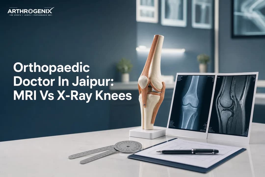
If you searched for Orthopaedic Doctor In Jaipur: MRI Vs X-Ray Knees, you likely want a clear, practical answer from a sports orthopaedic surgeon about which scan actually helps. In this guide, you will learn when an X-ray is enough, when an MRI adds value, and how we decide the next step for knee pain, swelling, or sports injury.
Why The Right Scan Matters For Knee Pain Decisions
First, imaging should answer a clinical question. Otherwise, it wastes time and money. So, a good orthopaedic surgeon in Jaipur starts with your story, a focused exam, and then chooses the most useful test.
In addition, knees often hurt for different reasons that feel similar. For example, early cartilage wear, a meniscus tear, and a ligament sprain can all cause pain on stairs. Therefore, the “best” scan depends on what we suspect after testing stability, joint-line tenderness, swelling, and range of motion.
Notably, imaging does not replace an exam. It supports it. As a result, you avoid two common problems: missing a serious injury or over-treating a harmless finding.
What We Commonly See In Clinic In Jaipur
In our day-to-day work at ArthroGenix, we see a repeating pattern. Many active people arrive with an MRI already done, yet they never had an X-ray. Meanwhile, some patients have multiple X-rays, but no one checked the meniscus or ACL clinically.
Because of this, we try to match symptoms to likely structures. Then we pick the scan that changes treatment. For instance, a locked knee after a twisting injury raises concern for a displaced meniscus tear, so MRI becomes more helpful. On the other hand, long-standing pain with bowing of the leg often needs an X-ray first to map arthritis severity.
Sports Orthopaedic Surgeon Guidance: MRI Vs X-Ray
Second, it helps to know what each test can and cannot “see.” X-rays show bones and joint alignment very well. However, MRI shows soft tissues like ligaments, meniscus, cartilage surfaces, and bone marrow changes.
Importantly, neither test is “better” in every case. Instead, each test answers different questions. Therefore, we often use them in sequence.
When An X-Ray Helps More Than You Think
An X-ray is quick and widely available. Moreover, it often gives the most direct answer for arthritis, fractures, alignment issues, loose bodies with calcification, and bone tumors suspected clinically. So, for pain that worsens with weight bearing and improves with rest, an X-ray can be the best starting point.
In addition, weight-bearing views matter. A standing X-ray can reveal joint-space narrowing that a non-weight-bearing film may miss. Therefore, we often request specific views instead of “any knee X-ray.”
According to the American College of Radiology Appropriateness Criteria, clinicians typically start with radiographs for chronic knee pain before advanced imaging. That approach improves diagnostic efficiency and avoids unnecessary MRI in many cases.
Common X-Ray Views We Request For Knees
- AP standing to assess joint space under load
- Lateral to review patella height and bony contour
- Skyline/Merchant to check patellofemoral arthritis and tracking
- Rosenberg view in selected arthritis cases
When MRI Is The Clear Winner
MRI becomes valuable when we suspect soft tissue injury. For example, ACL tear, PCL injury, meniscus tear, cartilage injury, or occult bone bruising often needs MRI to confirm severity and location. Consequently, MRI guides decisions on rehab versus surgery.
Similarly, persistent swelling after an injury can signal internal derangement. Therefore, if your knee feels unstable, gives way, or locks, MRI can reveal the mechanism. That information matters for athletes aiming to return to sport safely.
However, MRI can also show findings that are not the true pain source. In fact, many people have degenerative meniscus changes without symptoms. So, we interpret MRI in the context of your exam, not in isolation.
How We Decide Between MRI And X-Ray For Your Knee
Third, we use a simple decision framework. We clarify the injury timeline, the pain pattern, and the mechanical symptoms. Then we choose the test that will change treatment.
For a sports injury doctor Jaipur patients trust, the key is not “more imaging.” Instead, it is the right imaging at the right time. As a result, you get a faster path to recovery.
Here are practical scenarios that often decide the scan choice:
- Acute twist with a pop and swelling within hours: we examine for ACL injury and may order X-ray to rule fracture, then MRI for ligament assessment.
- Knee pain for months, worse on walking: we usually start with weight-bearing X-ray to check arthritis and alignment.
- Locked knee or cannot fully extend: MRI helps identify bucket-handle meniscus tear and urgency.
- Direct blow or fall with severe tenderness: X-ray first to evaluate fracture; MRI only if symptoms persist or we suspect hidden damage.
- Post-surgery pain or swelling: imaging depends on the procedure; sometimes X-ray assesses hardware, while MRI needs special planning.
Meanwhile, we also consider your age, sport, and goals. For instance, competitive athletes need clear return-to-play criteria. Therefore, MRI can support a safer plan when clinical uncertainty remains.
What The Reports Mean For Treatment Options
Finally, imaging should lead to an action plan. That plan may include physiotherapy, injection options, bracing, activity modification, or surgery. So, the real value is in connecting the report to your daily function.
At ArthroGenix, Dr Abhishek Saxena and the team focus on evidence-based pathways. Consequently, we often combine imaging with functional tests and rehab milestones. That approach suits active people as well as seniors with arthritis.
Below is how scan findings often map to treatment, in plain language:
- Arthritis on X-ray: start with targeted strengthening, weight management, and pain control options; consider knee replacement Jaipur planning only when symptoms and function justify it.
- ACL tear on MRI: decide between structured rehab and ACL reconstruction Jaipur based on instability, sport, and associated injury.
- PCL or multi-ligament injury: tailor bracing and rehab; consider PCL reconstruction Jaipur in select cases after detailed assessment.
- Meniscus tear: if mechanical locking persists, meniscus repair Jaipur or arthroscopic trimming may help; if degenerative, rehab often wins.
- Cartilage injury: joint preservation steps may include unloading, physiotherapy, and selected procedures; we match the option to size and location.
Importantly, imaging alone does not decide surgery. Your symptoms, exam findings, and activity needs matter just as much. Therefore, two people with the same MRI line item may need different care.
For example, a runner with true instability after an ACL tear may benefit from an ACL surgeon Jaipur consultation for reconstruction planning. On the other hand, a recreational walker with good stability may do well with rehab and guidance in sports medicine Jaipur programs.
For patient education on knee anatomy and imaging context, you can also review the MedlinePlus knee injuries and disorders resource. Additionally, for background on when MRI is used for musculoskeletal conditions, the NIAMS health topics pages offer reliable overviews.
If you want personalised next steps, explore ArthroGenix and see how our sports injury specialist approach blends diagnosis, minimally invasive treatment, and rehab planning. You can also visit our Jaipur sports orthopaedic clinic page to understand the evaluation flow before you book.
In short, the smartest path usually starts with a good clinical exam and the most appropriate scan, not the most expensive one. If you are comparing options after reading Orthopaedic Doctor In Jaipur: MRI Vs X-Ray Knees, a focused consult with a sports orthopaedic surgeon can clarify what your knee needs next. Results vary with injury pattern, fitness, and timing, so we keep recommendations transparent and tailored.